Characterization Equipment
In materials science, characterization helps to dissect a material’s physical and chemical structure. At the MILL, the Characterization Team uses multiple techniques to accomplish this, including optical and electron microscopy, optical profilometry, infrared spectroscopy, and various X-ray techniques (XRD & XRF). Note that X-ray techniques and SEMs require special X-ray safety training from GT.
The Characterization Team is led by Nicholas Stojanovic. Please contact Nick for more information!
If there is a specific piece of equipment you would like training on, please email the respective Technical Officer (TO) for that piece of equipment.
The list of Technical Officers can be found here.
Equipment List:
- Scanning Electron Microscopes (SEMs)
- Sputter Coater
- Digital Microscope
- Optical Profilometer
- Fourier-Transform Infrared Spectrometer (FTIR)
- X-ray Diffractometer (XRD)
- X-ray Fluorescence Spectrometer (XRF)
Scanning Electron Microscopes (SEMs)
Located in Main Lab (176)
SOPs: Phenom XL G2 SEM & Phenom ProX G5 SEM
Wiki: Scanning Electron Microscope (SEM)
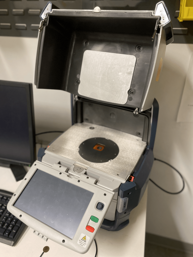
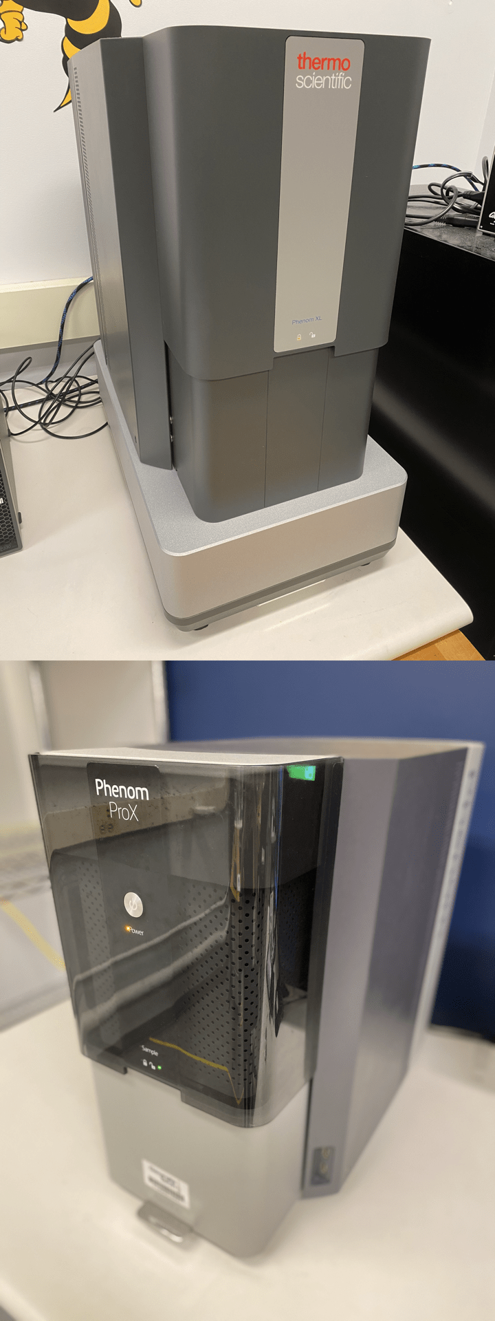
Both of our SEMs (scanning electron microscope) provide users with high-resolution electron microscopy images in just a few minutes. Both have maximum magnifications on the order of 100,000x with ~10 nm resolution. Samples must be thoroughly dried and degassed, and all loose particles must be blown free using compressed air. No magnetic samples are allowed.
The new Phenom XL G2 includes mixed secondary and backscattered electron detector imaging, returning to saved positions on your sample, and EDS/EDX (Energy-dispersive X-ray Spectroscopy) for quick elemental identification and EDS mapping. It also has the feature of ParticleMetric and PoroMetric for particle size and porosity measurements. The older Phenom ProX G5 includes a backscattered electron detector and can perform EDS/EDX (Energy-dispersive X-ray Spectroscopy) for quick elemental identification and EDS mapping. Two sample holders (for conductive and non-conductive samples, respectively) are available for use.
For more specifications, see our SOPs and Wiki page.
Sputter Coater
Located in Main Lab (176)
SOP: Cressington 108 Sputter Coater
Watch: Video tutorial for operating the Sputter Coater
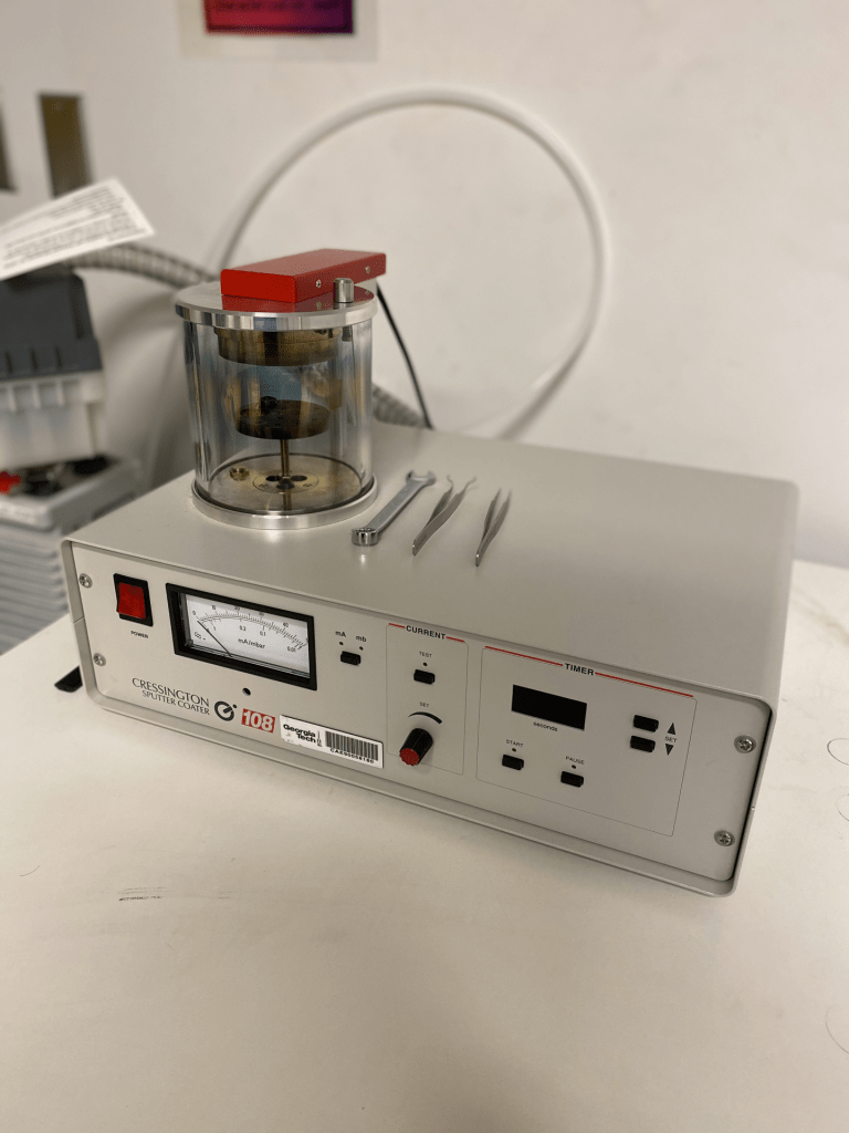
The Cressington 108 sputter coater deposits electrically conductive materials onto the desired surface to improve the surface’s electrical conductivity. Often, the MILL’s sputter coater is used in tandem with our SEMs because electron imaging requires a conductive surface. The MILL’s sputter coater primarily deposits gold, but other metal targets (Au:Pd, Pt, Pt:Pd) are also possible upon request. Additionally, the sputter coater serves as a test vacuum chamber for porous SEM samples which may undergo degassing.
For specifications, see our SOP and Wiki pages.
Digital Microscope
Located in Main Lab (176)
SOP: Leica DVM6 Digital Microscope
Wiki: Optical Microscopy
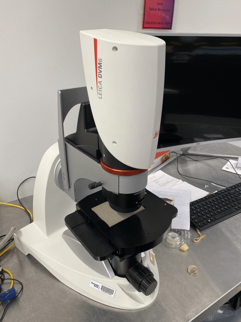
The digital microscope uses a digital camera unit combined with a powerful objective lens system to produce high-quality optical microscopy images down to a resolution of less than 1 µm at over 2000x magnification. This microscope has a motorized stage and focus drive more precise sample panning and focusing, and can automatically stitch images in the XY or layer images at different focal distances to form a Z-stack with enhanced depth of field. With this, it can also measure topographical features on the order of 10 µm.
Two objectives are available for the digital microscope (located below the microscope): PlanAPO FOV 12.55 and PlanAPO FOV 3.60.
For more specifications, see our SOP and Wiki pages.
Optical Profilometer
Located in Main Lab (176)
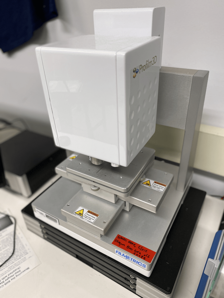
SOP: Filmetrics Profilm3D Optical Profilometer
Wiki: Optical Profilometer
The Profilm 3D Optical Profilometer uses white light interferometry to measure surface profiles and roughness. There are currently 10x, 20x, and 50x objectives that can resolve surface roughness down to 0.05 μm. From each image, ProFilm’s analysis software can generate analyzable topological maps, which can also be exported as .STL files for 3D printing.
For more specifications, see our SOP and Wiki pages.
Fourier-Transform Infrared Spectrometer (FTIR)
Located in Main Lab (176)
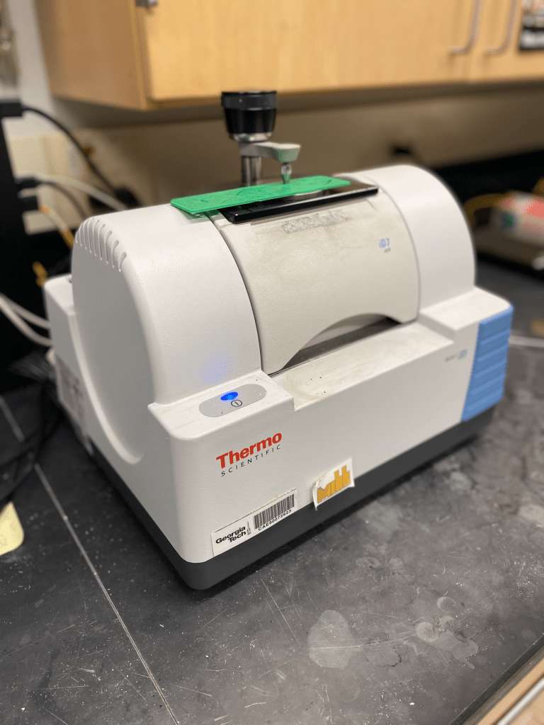
SOP: Nicolet iS5 FTIR
Wiki: Fourier-Transform Infrared Spectrometer (FTIR)
Fourier Transform Infrared Spectroscopy (FTIR) is a non-destructive characterization technique that uses infrared radiation to determine the molecular bonds present in organic material. These bonds must have a temporary or permanent dipole in order to be IR-active. When exposing the sample to infrared radiation, FTIR measures responses from a molecule’s vibrational modes, forming a molecular “fingerprint” used to identify chemical composition.
Our Nicolet iS5 FTIR has both transmission and ATR (attenuated total reflectance) accessories and is suitable for solid and liquid samples with minimal sample prep.
For more specifications, see our SOP and Wiki pages.
X-ray Diffractometer (XRD)
Located in X-ray Room (170)
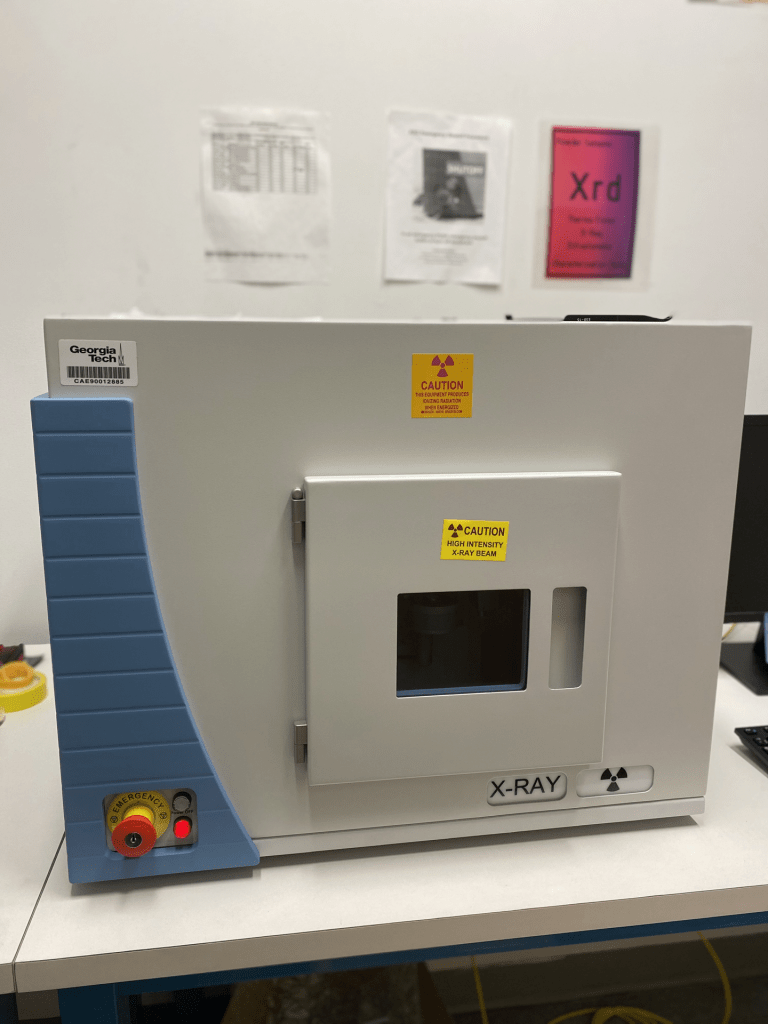
SOP: ARL Equinox 100 XRD
Wiki: X-ray Diffractometer (XRD)
X-ray diffractometry (XRD) detects crystalline phases in a sample by measuring the diffraction pattern of X-rays at varying angles. X-rays are used because the interplanar spacing for the vast majority of crystalline materials falls in the range of X-ray wavelengths (0.01-10 nm). Diffracted X-rays undergo either constructive or destructive interference by Bragg’s Law: nλ = 2d · sin(θ).
The Equinox XRD is set up for powder X-ray diffraction: for the best results, samples should be ground into a fine powder so that the collective group of crystals has a random orientation. It has a 2θ Range of 0° – 110° and uses a Cu Kα source (λ = 1.5406Å).
For more specifications, see our SOP and Wiki pages.
X-ray Fluorescence Spectrometer (XRF)
Located in X-ray Room (170)
SOP: Niton FXL XRF
Wiki: X-ray Fluorescence Spectrometer (XRF)
X-ray fluorescence spectroscopy (XRF), also known as X-ray emission spectroscopy (XES), is a non-destructive characterization technique that gives elemental composition information. Our XRF is a field unit, allowing for minimal sample prep and fast readings in exchange for slightly lower resolution. It can detect elements from magnesium onward (Z ≥ 12) on the periodic table and is best suited for ceramic and metallic samples. The Niton FXL XRF accepts bulk, powdered, and liquid samples.
For more specifications, see our SOP and Wiki pages.